Work / VMI
Velocity Map Imaging (VMI)
In 1997, Eppink and Parker revolutionized electron/ion imaging with their innovative extension of Chandler and Houston’s earlier technique. The Velocity Map Imaging (VMI) technique allowed for exquisite mapping of charged species based on their kinetic energies, with an electrostatic potential projecting them onto concentric rings of unique radii. This sophisticated spectrometer had a 4π solid angle collection efficiency and was versatile, doubling as a time-of-flight mass spectrometer and photoelectron imaging spectrometer.
The VMI spectrometer was a marvel of engineering, composed of three regions that worked in tandem to capture the movements of charged species. The ionization region, the site of the light-matter interaction, housed extractor and repeller plates held at high potentials for complete collection. The focusing region featured extractor and focusing electrodes, which enabled electrostatic focusing and precise resolution control. Finally, the drift region allowed for smooth spatial and temporal resolution before the charged species were collected by a two-dimensional detector.
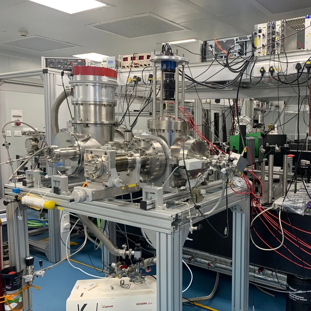
Our homemade VMI spectrometer, the Thick Lens Velocity Map Imaging (TL-VMI) spectrometer, was a reflection of the artistic and academic endeavor to capture the beauty and intricacies of charged species. This nine-plate system promised finer resolution and a more in-depth understanding of the movements of charged particles. The simulation, construction, and assembly of this instrument were a testament to the art and science of imaging techniques, pushing the boundaries of knowledge and understanding of the world around us.